Dermpath Diagnosis
Pretibial Myxedema
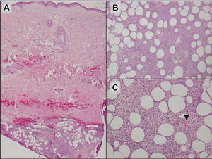
Pretibial myxedema is a cutaneous mucinosis associated with thyroid dysfunction. The differential diagnosis includes nephrogenic systemic fibrosis...
Pretibial myxedema is a cutaneous mucinosis associated with thyroid dysfunction. The differential diagnosis includes nephrogenic systemic fibrosis...
Hailey-Hailey disease typically presents as suprabasal blisters with a perivascular and interstitial lymphocytic infiltrate. The differential...

Giant cell tumors of soft tissue present as multilobulated masses in the skin or subcutaneous tissue, most commonly appearing on the trunk, upper...