Photo Challenge

Punctate Depigmented Macules

Author and Disclosure Information


An otherwise healthy 54-year-old black man presented with a 10-year history of spotty pigmentary loss in a band on the left side of the abdomen, flank, and back. He denied a history of rash or inflammation in the area and had not experienced confluent depigmentation. He reported that initially he had only a few “white dots,” and over the next 5 to 7 years, he developed more of them confined within the same area. On presentation, he stated new areas of depigmentation had not developed in several years. The band was completely asymptomatic and had not been treated with any prescription or over-the-counter medications. On examination he had multiple 2- to 3-mm confettilike depigmented macules that seemed to be centered around follicles in a band with blaschkoid distribution extending across the left side of the abdomen, flank, and back. The band did not cross the midline and similar lesions were not present elsewhere. A punch biopsy of one of the depigmented macules revealed a markedly diminished number of melanocytes along the junction as well as a decrease in melanin, which was confirmed by Melan-A and Fontana stains, respectively.

What's the diagnosis?

a. blaschkoid punctate vitiligo

b. chemical leukoderma

c. hypopigmented mycosis fungoides

d. postinflammatory pigment alteration

e. progressive macular hypomelanosis

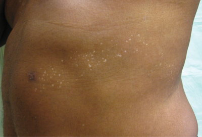
The Diagnosis: Blaschkoid Punctate Vitiligo

Based on the patient’s clinical appearance as well as the histologic findings, the diagnosis of vitiligo was made. Although vitiligo is certainly not uncommon and punctate vitiligo is a known clinical presentation,1 punctate vitiliginous depigmentation conforming to lines of Blaschko is unique. Follicular repigmentation in a patch of vitiligo potentially could lead to this “spotty” appearance, but our patient maintained that the band was never confluently depigmented and that small macules arose within normally pigmented skin. The patient’s adult age at onset makes this case even more unusual.

Follicular repigmentation in vitiligo is fairly well understood, as the perifollicular pigment is formed by upward migration of activated melanoblasts in the outer root sheath.2 Follicular depigmentation as well as selective or initial loss of melanocytes around hair follicles in early vitiligo has not been described. It is unclear if the seemingly folliculocentric nature of the patient’s vitiliginous macules was a false observation, coincidental, or actually related to selective melanocyte loss around follicles.

Blaschkoid distribution has been described in numerous skin disorders and is known to be based on genetic mosaicism.3 Most of these disorders are X-linked and/or congenital. However, many acquired skin conditions have been described exhibiting blaschkoid distribution, such as vitiligo, psoriasis, lichen planus, atopic dermatitis, and mycosis fungoides.4,5

Confettilike depigmentation has been described as an unusual clinical variant of vitiligo.1 It also has been reported after psoralen plus UVA therapy in patients with more classic vitiligo,6 numerous domestic chemicals,7 and in association with mycosis fungoides.8 In these cases, punctate lesions were disseminated, symmetric on extremities, or limited to areas exposed to chemicals.

Recommended Reading

Ashiness
MDedge Dermatology
Hair washing – Too much or too little?
MDedge Dermatology
AAD 2014 sessions offer something for everyone
MDedge Dermatology
VIDEO: Vitiligo gene hunt could open door to eventual drug treatment
MDedge Dermatology
Pulsed Dye Laser for the Treatment of Macular Amyloidosis: A Case Report
MDedge Dermatology
The Skinny Podcast: Vitiligo research update, Dr. Rockoff on puzzling prescription rules, and the latest dermatology headlines
MDedge Dermatology
Discrepancies in dyschromia
MDedge Dermatology
Chlorpromazine-Induced Skin Pigmentation With Corneal and Lens Opacities
MDedge Dermatology
Generalized Yellow Discoloration of the Skin
MDedge Dermatology
The springtime eruptions
MDedge Dermatology