Photo Challenge

Crusted Plaque in the Umbilicus

Author and Disclosure Information

A 74-year-old man presented to our outpatient dermatology clinic with an asymptomatic umbilical lesion of unknown duration. The patient believed the lesion was a scar resulting from a prior laparoscopic repair of an umbilical hernia. However, the patient reported epigastric abdominal pain and diarrhea of 1 month's duration that he believed was due to the stomach flu. The patient denied fever, chills, loss of appetite, or weight loss. History was remarkable for hypertension, hyperlipidemia, coronary artery disease, chronic kidney disease, and emphysema. The patient had a surgical history of percutaneous transluminal coronary angioplasty in addition to the laparoscopic umbilical hernia repair. The patient's medications included pantoprazole, ondansetron, diphenoxylate-atropine as needed, amlodipine, lisinopril-hydrochlorothiazide, simvastatin, and aspirin. Physical examination revealed a 1×2-cm pink, nodular, firm plaque with crust at the umbilicus that was tender on palpation. A shave biopsy of the umbilicus was performed and sent for both pathological and immunohistochemical analysis.

What's the diagnosis?

nonmelanoma skin cancer

psoriasis

pyogenic granuloma

scar

Sister Mary Joseph nodule

The use of dermoscopy has been advocated as an adjuvant tool in delineating benign and malignant umbilical lesions when an atypical polymorphous vascular pattern indicating neovascularization has been observed with neoplastic growth.5 Once a suspicious umbilical lesion is identified, the first step should be to obtain a skin biopsy or to use fine needle aspiration for cytology.6 Biopsy is especially relevant in the background of cancer history because SMJN may present with cancer recurrence.3 Once one of these is obtained, histological and immunohistochemical analysis will guide further workup and diagnosis of the umbilical lesion.

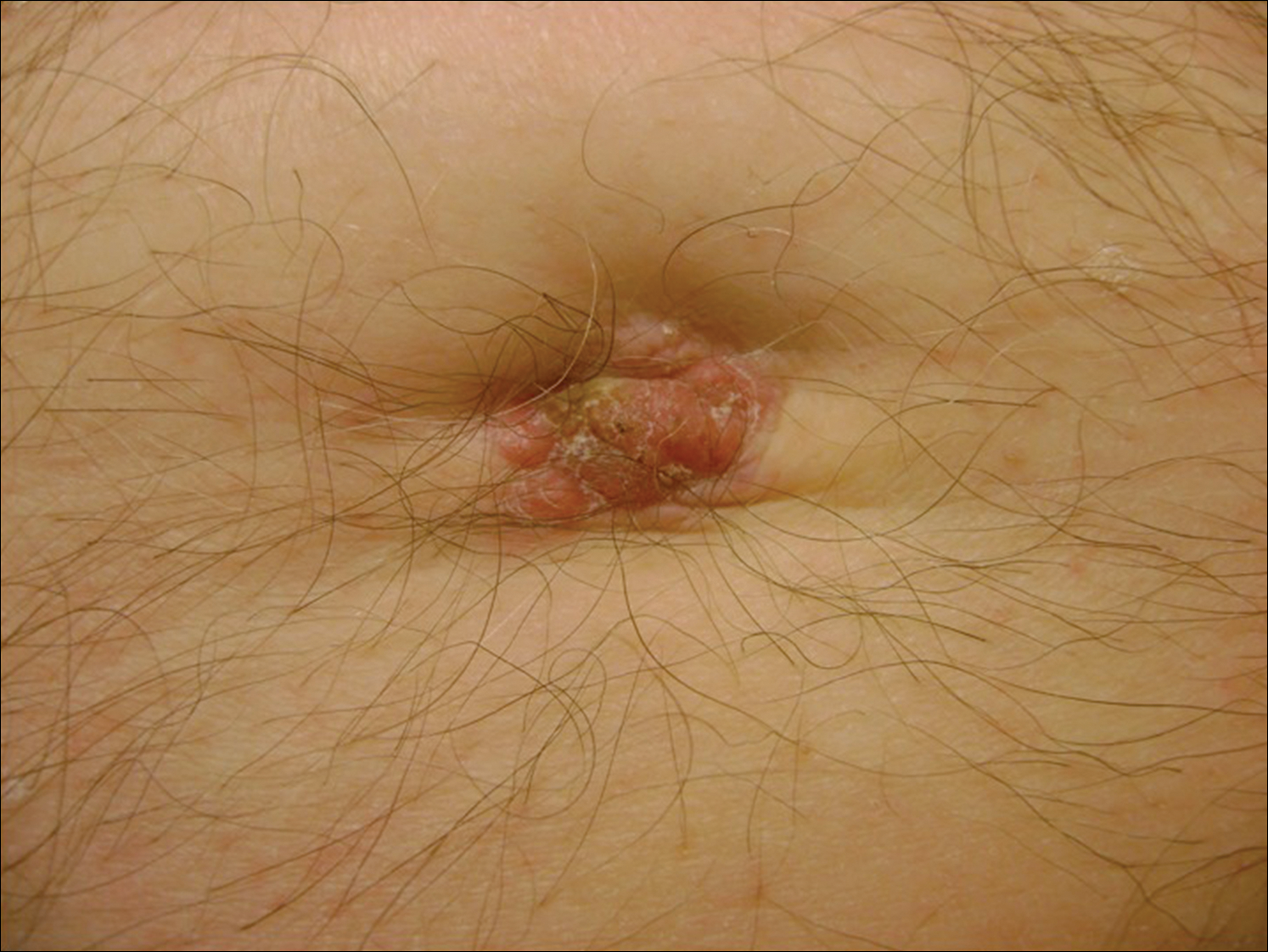
The importance of reviewing such cases lies in the variable presentation of cutaneous metastases such as SMJN and the grim prognosis that accompanies this finding. It presents as a firm indurated plaque or nodule that may present with systemic symptoms suggestive of malignancy, though in 30% of cases it is the sole initial sign.7 The nodule may be painful if ulcerated or fissured. Bloody, serous, or purulent discharge may be present. After diagnosis of an SMJN, most patients succumb to the disease within 12 months. Thus, it is vital for dermatologists to investigate umbilical lesions with great caution and a high index of suspicion.

Pages

Recommended Reading

Painful Ulcerations Above the Malleoli
MDedge Dermatology
Desmoplastic Hairless Hypopigmented Nevus
MDedge Dermatology
Metastatic Crohn Disease Clinically Reminiscent of Erythema Nodosum on the Right Leg
MDedge Dermatology
Development of Bullous Pemphigoid in a Patient With Psoriasis and Metabolic Syndrome
MDedge Dermatology
The Proposed Rule and Payments for 2017: The Good, the Bad, and the Ugly
MDedge Dermatology
Pruritic Papules on the Scalp and Arms
MDedge Dermatology
Bullous Pemphigoid Associated With a Lymphoepithelial Cyst of the Pancreas
MDedge Dermatology
Diagnosis of a Rapidly Growing Preauricular Nodule: Chondroid Syringoma or Pleomorphic Adenoma?
MDedge Dermatology
Epidermodysplasia Verruciformis and the Risk for Malignancy
MDedge Dermatology
Blaschkoid Unilateral Patch on the Chest
MDedge Dermatology