DermaDiagnosis
Thinking Pimple? That’s Too Simple
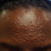
A 13-year-old girl presents with stubborn forehead lesions that were previously diagnosed as acne. Any attempts at treatment thus far have been...

A 5-year-old boy is referred to dermatology for evaluation of recent color changes to the skin around a congenital lesion. Located on his mid low back, the polygonal lesion measures 8 x 5 cm and is uniformly dark brown with a mammillated, hair-bearing surface. The plaque has grown proportionately with the child but otherwise remained stable.
A year ago, however, the patient’s family noticed that the normal brown skin around the lesion was turning white. This “halo” became noticeably larger over the span of the year—effectively doubling the size of the lesion.
The child’s type V skin is in sharp contrast to the porcelain white band that parallels the margins of his lesion. The surface of the depigmented skin is completely smooth, with no epidermal changes. Faint but definite depigmentation is noted on the periocular skin of both eyes, in addition to well-defined depigmentation on his fingertips and the perionychial areas of all 10 fingers.
The family asserts that the boy is otherwise healthy and that there is no family history of similar phenomena. The rest of the examination is unremarkable.
A 13-year-old girl presents with stubborn forehead lesions that were previously diagnosed as acne. Any attempts at treatment thus far have been...
A 15-year-old boy is embarrassed by persistent depigmentation that leaves the skin around his nose gray and scaly. Can you help by providing a...

A 12-year-old African-American girl had lesions on her trunk since shortly after birth. Her pediatrician was confident that she would "outgrow"...